“Ten hours in. My eyes hurt.”

After a long coding session, dry, tired eyes are what you feel. What you can’t feel happens beneath the surface, to cells that can’t be replaced, and this matters for your long-term eye health. Most coders don’t think about it enough.

Locked in for hours. Eyes dry.

After a long coding session, dry, tired eyes are what you feel. What you can’t feel happens beneath the surface, to cells that can’t be replaced, and this matters for your long-term eye health. Most coders don’t think about it enough.

Your theme isn’t the problem

After a long coding session, dry, tired eyes are what you feel. What you can’t feel happens beneath the surface, to cells that can’t be replaced, and this matters for your long-term eye health. Most coders don’t think about it enough.

A programmer's desk late in the evening: a design document on one monitor, a coding assistant's terminal session on the other, code open on a laptop, the chair empty.

33 sources · updated 11 Nov 2026 · by Clearer®

Locked in, you blink less and the eyes dry out

After a long session the eyes feel dry, sore, sometimes burning, and an ache builds behind them from holding focus. That dryness is the surface: you blink less, and more of your blinks stop halfway, so the tear film breaks up and the eye starts to dry out.

“I program for more than 8 hours a day and have started to feel eye irritation more often”

A post in r/learnprogramming, November 2024

“I’ve found that my eyes end up feeling very dry throughout the day because I’m looking at a monitor and staring at code.”

A post in r/cscareerquestions, April 2017

“…my eyes can get irritated as little as 20 mins into a work sesh”

A post in r/webdev, October 2024

But that’s not the whole story. Dryness is more than a lack of moisture: as a broken tear film evaporates, what is left turns saltier, injuring the surface cells and switching on inflammation. Inside those cells oxidative stress builds as well, from reactive forms of oxygen that damage fats and proteins; in 80 young adults, the reactive oxygen in them rose over four hours on a screen.

Half-blinks a minute on a phone; more is worse

6 half-blinks a minute in minute 1, 15 in minute 60.

Typical counts for 12 adults aged 18–23, reading on a phone for an hour in a lab.
Figure 1: reactive oxygen at the eye's surface over four hoursLine chart. Change in reactive oxygen in surface cells of the eye from the start, at 0, 1 and 4 hours. Phone group (50 people): 0, +12.5%, +31.2%. Computer-screen group (30 people): 0, +8.9%, +13.2%. Higher is worse.Reactive oxygen at the eye's surface, change from the start; higher is worse+10%+20%+30%+40%00 h1 h4 h+31%Phone+13%Computer
Figure 1: reactive oxygen at the eye's surface over four hoursLine chart. Change in reactive oxygen in surface cells of the eye from the start, at 0, 1 and 4 hours. Phone group (50 people): 0, +12.5%, +31.2%. Computer-screen group (30 people): 0, +8.9%, +13.2%. Higher is worse.Reactive oxygen at the eye's surface,change from the start; higher is worse+10%+20%+30%+40%00 h1 h4 h+31%Phone+13%Computer
Figure 1. Follow the two lines past the first hour: reactive oxygen at the eye’s surface keeps climbing on both screens, and climbs further on the phone.
Illustration of the tear film: a thin golden oil layer over clear water, over a mucus layer on the surface cells.
The tear film: oil over water over mucus
Illustration of a cell membrane seen side-on: a double layer of fats, with yellow pigment molecules among the tails.
A cell membrane: fats that reactive oxygen can damage
MeasureValueWho
Lipid-damage marker 4-HNE in tearsabout 10× higher with dry eye44 people with dry eye, 33 without

Reactive oxygen was measured with a fluorescent dye in surface cells (conjunctiva), before and after a puzzle game at 80% brightness on a 5.1-inch phone (50 people) or a 19-inch computer screen (30 people); the two groups were not randomised. Tear 4-HNE was measured in people already diagnosed with dry eye.

What it adds up to: reactive oxygen rising over hours on a screen, and, in people with dry eye, damaged fats in the tears and inflammation at the surface.

Drops soothe that surface and are worth keeping, but reviews of dry eye find that extra moisture doesn’t address the inflammation and oxidative stress beneath it; to hold reactive oxygen in check, the surface’s cells rely on antioxidants.

Every cell makes reactive oxygen as it uses oxygen, and the surface’s cells are no exception. More importantly, the retina is under oxidative stress too, from a metabolic load that never lets up, and none of it is felt.

The retina uses oxygen faster than the brain, in cells never replaced

Figure 2a: where it happensIllustration: the eye seen from above, light entering through the cornea and lens to the macula at the centre of the retina, marked as a yellow spot and ringed, with the optic nerve leaving on the nasal side. The tear film on the front surface, where dryness is felt, is a blue crescent with a few reactive-oxygen dots on it, as measured there; the retina lining the back, where nothing is felt, is a pale rose band. Below, one rod from the macula: its inner segment packed with mitochondria and its outer segment a stack of membrane discs, ringed, which part c enlarges.aEye, seen from aboveLightOptic nerveMacula, the centre of the retinaDryness is felt hereNothing is felt hereOne rodInner segment:packed withmitochondriaStacked membranediscs: see c
Figure 2a: where it happensIllustration: the eye seen from above, light entering through the cornea and lens to the macula at the centre of the retina, marked as a yellow spot and ringed, with the optic nerve leaving on the nasal side. The tear film on the front surface, where dryness is felt, is a blue crescent with a few reactive-oxygen dots on it, as measured there; the retina lining the back, where nothing is felt, is a pale rose band. Below, one rod from the macula: its inner segment packed with mitochondria and its outer segment a stack of membrane discs, ringed, which part c enlarges.aEye, seen from aboveLightOptic nerveMacula, the centre of the retinaDryness is felt hereNothing is felt hereOne rodInner segment: packed withmitochondriaStacked membrane discs: see c
Figure 2a. Dryness is felt on the surface at the front. The retina lines the back of the eye, where the cells that catch light sit; the chemistry there is not felt.
  • They work all day

    Rods work hardest in the dark, and cones keep working through daylight.

  • Their fat oxidises easily

    About a third to over half of the fatty acids in their light-catching discs are DHA, the fat that oxidises most easily.

  • They aren’t replaced

    Lost ones are not replaced: in donor eyes aged 27 to 90, rod density in the central retina fell by about 30% across adult life.

Figure 2b: the chainIllustration: one half of the disc membrane, closer. A radical (R) takes a hydrogen from a DHA tail (1); the damaged fat takes oxygen (2) and takes a hydrogen from the next fat (3), which does the same to the next. Three fats are damaged in a row, marked in the damage colour. Vitamin E, ringed, gives its hydrogen to the radical and the chain stops there (4); the fat beyond it is still intact.bThe chain, step by stepR123334Damaged fatsStill intact1A radical takes a hydrogenL•2The damaged fat takes oxygenLOO•3It takes a hydrogen from the nextfatLOOH4Vitamin E stops the chain
Figure 2b: the chainIllustration: one half of the disc membrane, closer. A radical (R) takes a hydrogen from a DHA tail (1); the damaged fat takes oxygen (2) and takes a hydrogen from the next fat (3), which does the same to the next. Three fats are damaged in a row, marked in the damage colour. Vitamin E, ringed, gives its hydrogen to the radical and the chain stops there (4); the fat beyond it is still intact.bThe chain, step by stepR123334Damaged fatsStill intact1A radical takes a hydrogenL•2The damaged fat takes oxygenLOO•3It takes a hydrogen from the next fatLOOH4Vitamin E stops the chain
In lab and animal studies, intense light and oxygen can start a chain reaction in the retina’s delicate fats, one damaged fat damaging the next. Figure 2b. One damaged fat takes a hydrogen from the next, link by link through the disc’s membrane, until vitamin E stops it.

The cells that catch light are the retina’s photoreceptors; at the back of the eye, they turn light into sight.

A radical is a molecule with an unpaired electron. One can damage fat after fat, and the chain reaction leaves fragments which stick to the cells’ proteins.

Where the reactive oxygen comes from: mitochondria occasionally pass a single electron to oxygen, making superoxide; an enzyme turns it into hydrogen peroxide, and where free iron is present that can become the hydroxyl radical, the most reactive of all. A little leaks all the time; that leak can start the chain. Light adds a second source: in lab studies, spent visual pigment and the age pigment lipofuscin absorb violet-blue light and pass the energy to oxygen.

The rod’s energy is spent as ATP, the cell’s fuel; the retina’s oxygen use is “one of the highest… of any tissue in the body, exceeding that of the brain” (Uddin 2025).

Why screens don’t add to the load: in monkeys, light lowered the photoreceptors’ oxygen use by 16 to 36%, so the peak load is in the dark. Seeing at all is the cost.

DHA’s share depends on species and method: 32% of fatty acids in photoreceptor outer segments (30–35% in human donor eyes); at least 50% and up to 60% in rod outer-segment membranes in other papers.

The chain has a name, lipid peroxidation. A radical tears a hydrogen off one DHA chain; that fat, now a radical itself, seizes oxygen and does the same to the next. DHA’s six double bonds leave the hydrogens a radical pulls off, which is why it oxidises more easily than any common fat. The fragments include 4-HHE (from DHA), 4-HNE (from omega-6 fats) and MDA.

A developer's desk by daylight: a monitor raised on an arm with a light bar, code in large type in two panes, an ergonomic chair.

Your setup tunes the screen. None of it looks after the retina.

FixWhat it changes
Done: Bigger text, zoomFewer lost blinks: in one lab study, blinking fell least at 330% zoom
Done: Theme, light or darkLegibility, to taste; no study shows either protects the eyes
Done: BreaksRest for focussing, the main source of the ache behind the eyes
Done: DropsWater for the tear film
Not covered: The retinaNone of the above. Its defences depend on what you eat.

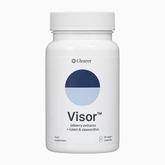
Introducing Visor™ for long screen days, today and for the decades ahead

Targeted nutrients for people with long screen time, each researched for the surface that dries out by evening or for the retina behind it, which has to last for decades.

  1. Drops soothe it for a while. In a six-month trial in heavy screen users, tear break-up time rose with lutein and zeaxanthin and not on placebo.

  2. Lutein and zeaxanthin form a natural light filter at the centre of the retina, a yellow layer that absorbs blue light before it reaches the cells that catch light. Your body can’t make either.

  3. Vitamin E stops the chain in the retina’s delicate fats. Vitamin C restores it, so it can stop the next.

  4. The body turns it into vitamin A, which the cells that catch light use to turn light into sight.

The screen-dried surface

Drops soothe it for a while. In a six-month trial in heavy screen users, tear break-up time rose with lutein and zeaxanthin and not on placebo.

No hype. Every ingredient, and what it does.

The light filter

13 mg together, about seven to nine times a typical US diet.

IngredientOn the labelWhat it does
Lutein12 mgBuilds the yellow pigment at the centre of the retina, which absorbs blue light
Zeaxanthin1 mgJoins the pigment; quenches reactive oxygen

Vitamins E and C, as a team

Alone, vitamin E isn’t enough: used up, in lab tests it can keep the oxidation going.

IngredientOn the labelWhat it does
Vitamin E20 mgStops the peroxidation chain in the membrane
Vitamin C80 mgRestores spent vitamin E
Vitamin B21.4 mgBecomes FAD, which keeps glutathione recharging

Beta-carotene into vitamin A

IngredientOn the labelWhat it does
Beta-carotene4.8 mgBecomes vitamin A, which the cells that catch light use to turn light into sight

Boost it with bilberry extract

IngredientOn the labelWhat it does
Bilberry extract90:1, 25% anthocyaninsAnthocyanins, studied in people after screen work

Figure 2c: what is in the disc membraneIllustration: one disc membrane at molecular scale, water above and below. Each fat (phospholipid) has a straight saturated tail and a DHA tail bent at each of its six double bonds. Vitamin E, ringed, lies among the tails with its ring at the surface. Lutein stands across the membrane, one end at each surface. Rhodopsin crosses the membrane, holding vitamin A (11-cis-retinal), which the body makes from beta-carotene.cDisc membrane, molecularVitamin E: head at thesurface, tail among the fatsDHA: sixdouble bondsLutein and zeaxanthin:span the membraneRhodopsin, holdingvitamin A(11-cis-retinal)vitamin ABeta-carotene
Figure 2c: what is in the disc membraneIllustration: one disc membrane at molecular scale, water above and below. Each fat (phospholipid) has a straight saturated tail and a DHA tail bent at each of its six double bonds. Vitamin E, ringed, lies among the tails with its ring at the surface. Lutein stands across the membrane, one end at each surface. Rhodopsin crosses the membrane, holding vitamin A (11-cis-retinal), which the body makes from beta-carotene.cDisc membrane, molecularVitamin E: head at the surface, tailamong the fatsDHA: six double bondsLutein and zeaxanthin: span themembraneRhodopsin, holding vitamin A(11-cis-retinal)vitamin ABeta-carotene

Vitamin E reacts with the chain-carrying radical “almost four orders of magnitude faster” than that radical’s attack on the next fat, both estimated for biological membranes (Bakker 2024).

The relay: vitamin C restores spent vitamin E; glutathione, the cell’s own antioxidant, restores vitamin C; and glutathione reductase, which recharges glutathione, runs on FAD, made from vitamin B2.

Figure 2d: the relayIllustration: a ladder from the membrane surface out into the water. Spent vitamin E at the surface is restored by vitamin C, which becomes dehydroascorbate; glutathione (GSH) restores vitamin C and becomes GSSG; glutathione reductase, running on FAD made from vitamin B2, recharges glutathione.dThe relay that keeps vitamin EworkingSpent vitamin Ein the membraneVitamin C restores EVitamin Cnow spent(dehydroascorbate)Glutathione restores CGSHGlutathione (GSH)now spent(GSSG)recharges glutathioneFADGlutathione reductaseruns on FAD, made from vitamin B2
Figure 2d: the relayIllustration: a ladder from the membrane surface out into the water. Spent vitamin E at the surface is restored by vitamin C, which becomes dehydroascorbate; glutathione (GSH) restores vitamin C and becomes GSSG; glutathione reductase, running on FAD made from vitamin B2, recharges glutathione.dThe relay that keeps vitamin E workingSpent vitamin Ein the membraneVitamin C restores EVitamin Cnow spent(dehydroascorbate)Glutathione restores CGSHGlutathione (GSH)now spent(GSSG)recharges glutathioneFADGlutathione reductaseruns on FAD, made from vitamin B2

In retinal tissue, vitamin C restored vitamin E and glutathione alone did not; the glutathione-to-vitamin C step is described for cells in general.

The macular pigment absorbs most strongly at about 460 nm. Each photon flips one vitamin A molecule (11-cis-retinal, to all-trans) in the visual pigment, and the visual cycle rebuilds it.

A typical US diet supplies 1.5 to 1.8 mg of lutein and zeaxanthin a day.

The body makes vitamin A from beta-carotene only as it needs it.

Vitamin B2 keeps the relay behind vitamin C running: it becomes FAD, which glutathione reductase needs.

The 24 mg in the first trial below was about 20 mg lutein, 2.4 mg zeaxanthin and 1.7 mg meso-zeaxanthin.

What six months of lutein changed in heavy screen users

Randomised and placebo-controlled, each dose shown beside Visor’s.

Eye strain

After six months, people reported less eye strain and fewer headaches than on placebo, and recovered faster from a bright flash. Their macular pigment, the eye’s own blue-absorbing layer, grew denser.

Who
48 young adults on screens at least 6 hours a day
How long
6 months
Dose tested
24 mg a day of lutein, zeaxanthin and meso-zeaxanthin · Visor™: 12 mg lutein, 1 mg zeaxanthin
Stringham et al., Foods 2017 (opens in a new tab)
Tear film

Tear break-up time rose in the lutein and zeaxanthin group and not on placebo. Break-up time is how long the tear film holds between blinks, one of the standard tests for dry eye.

Who
70 adults aged 18 to 65 on screens more than 6 hours a day
How long
6 months
Dose tested
10 mg lutein, 2 mg zeaxanthin a day · Visor™: 12 mg lutein, 1 mg zeaxanthin
Lopresti & Smith, Frontiers in Nutrition 2025 (opens in a new tab)

Both trials tested Visor’s ingredients at their own doses, not Visor™ itself. Visor™ adds what neither tested: vitamins E, C and B2.

Start with six months, the length of both trials

Blood lutein starts rising within two weeks, and the macular pigment builds over the months after. Both screen-user trials ran six months.

Six bottles of Visor™, the six-month bundle.
Choose a plan
Guarantee
180 days, money back: email info@clearereyes.com with your order number for a full refund.

The sceptic’s questions, answered

Because Visor™ is made for one kind of day: long hours on a screen.

A targeted antioxidant system for screen time. Lutein and zeaxanthin build the retina’s light filter, and vitamins E and C work as a team, each chosen for people who spend most of a working day on screens.

Research-backed doses. Its 12 mg of lutein sits between the lutein doses of the two six-month trials in heavy screen users: 10 mg in one, about 20 mg in the other.

Clean and bioavailable. Lutein, zeaxanthin and beta-carotene dissolve in fat, so taken with a meal they’re absorbed with it, and the body turns beta-carotene into vitamin A only as it needs it.

No study shows that dark mode, or light mode, protects the eyes. Use whichever reads better for you; bigger text and regular breaks are the supported habits, and neither theme reaches the chemistry in the retina.

Partly. Lutein and zeaxanthin form a natural light filter at the centre of the retina, a yellow layer that absorbs blue light before it reaches the cells that catch light. Glasses and night modes change the light outside the eye, and they’re worth keeping; the filter and the vitamins in Visor™ work inside it.

Visor™ gives 13 mg of lutein and zeaxanthin together. The two screen-user trials gave 12 mg and 24 mg of the two, and a typical US diet supplies 1.5 to 1.8 mg, so Visor™ gives about seven to nine times that.

Not the next morning: it’s upkeep, like the rest of your setup. Both trials read their results at six months, and the cells it’s for aren’t replaced once lost, so the upkeep starts now.

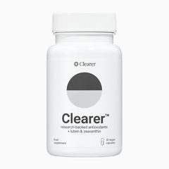
Yes. Clearer® is a different formula, and together they give about 22 mg of lutein and 160 mg of vitamin C a day. What Visor™ adds is vitamin E, vitamin B2 and beta-carotene.

The sources behind this page

  1. Golebiowski B et al. Curr Eye Res 2020. One hour of smartphone reading. PMID 31573824 (opens in a new tab)
  2. Argilés M et al. IOVS 2015. Blink rate across reading platforms. PMID 26517404 (opens in a new tab)
  3. Bron AJ et al. TFOS DEWS II Pathophysiology Report. Ocul Surf 2017. PMID 28736340 (opens in a new tab)
  4. Choi JH et al. PLoS One 2018. Smartphone use and reactive oxygen at the ocular surface. PMID 30379901 (opens in a new tab)
  5. Choi W et al. Curr Eye Res 2016. Lipid-peroxidation markers in dry-eye tears. PMID 26731289 (opens in a new tab)
  6. Wolffsohn JS et al. TFOS Lifestyle: the digital environment. Ocul Surf 2023. PMID 37062428 (opens in a new tab)
  7. Kamøy B et al. Acta Ophthalmol 2022. Screen use and dry eye: preventive measures (review). PMID 35122403 (opens in a new tab)
  8. Okawa H et al. Curr Biol 2008. Photoreceptor energy use. PMID 19084410 (opens in a new tab)
  9. Uddin MI. Prog Retin Eye Res 2025. Retinal oxygen, from microelectrodes to imaging (review). PMC13283042 (opens in a new tab)
  10. Ahmed J et al. IOVS 1993. Oxygen distribution in the macaque retina (light lowers photoreceptor oxygen use). PMID 8449672 (opens in a new tab)
  11. Murphy MP. Biochem J 2009. How mitochondria produce reactive oxygen species. PMID 19061483 (opens in a new tab)
  12. Bakker LM, Boulton ME, Różanowska MB. Antioxidants 2024. Partly oxidised DHA and cultured retinal cells (its introduction reviews DHA in the retina). PMID 39594569 (opens in a new tab)
  13. Ayala A, Muñoz MF, Argüelles S. Oxid Med Cell Longev 2014. Lipid peroxidation. PMID 24999379 (opens in a new tab)
  14. Leng X et al. Biophys J 2015. Vitamin E in DHA-rich membranes. PMID 26488652 (opens in a new tab)
  15. Bowry VW, Ingold KU, Stocker R. Biochem J 1992. Vitamin E without a co-antioxidant. PMID 1463440 (opens in a new tab)
  16. Stoyanovsky DA et al. Curr Eye Res 1995. Vitamin C restores vitamin E in retinal tissue. PMID 7796601 (opens in a new tab)
  17. Sinha T et al. Front Cell Dev Biol 2020. Flavins (vitamin B2) in the retina. PMC7481326 (opens in a new tab)
  18. Linus Pauling Institute. Vitamin E. lpi.oregonstate.edu (opens in a new tab)
  19. Jain S et al. Sci Rep 2023. Macular pigment and sensitivity to glare. PMID 37899478 (opens in a new tab)
  20. Widomska J, Subczynski WK. J Clin Exp Ophthalmol 2014. Lutein and zeaxanthin in membranes. PMID 24883226 (opens in a new tab)
  21. USDA ARS, What We Eat in America, NHANES 2017–March 2020, Table 1 (lutein and zeaxanthin intake, adults 20 and over). ars.usda.gov (opens in a new tab)
  22. Linus Pauling Institute. Vitamin A. lpi.oregonstate.edu (opens in a new tab)
  23. Ramkumar S et al. J Lipid Res 2021. Regulated conversion of beta-carotene to vitamin A. PMC8010212 (opens in a new tab)
  24. Sanges D et al. J Clin Invest 2016. Lost retinal neurons are not replaced. PMID 27427986 (opens in a new tab)
  25. Curcio CA et al. IOVS 1993. Ageing of the human photoreceptor mosaic. PMID 8225863 (opens in a new tab)
  26. Stringham JM, Stringham NT, O’Brien KJ. Foods 2017. PMID 28661438 (opens in a new tab)
  27. Lopresti AL, Smith SJ. Front Nutr 2025. PMID 39963662 (opens in a new tab)
  28. Stringham JM, Stringham NT. Exp Eye Res 2016. Timing of lutein in blood and macular pigment. PMID 27426932 (opens in a new tab)
  29. Kosehira M et al. Nutrients 2020. Bilberry extract after screen work. PMC7146147 (opens in a new tab)
  30. Sheppard AL, Wolffsohn JS. BMJ Open Ophthalmol 2018. Digital eye strain: focusing stress and dry eye. PMID 29963645 (opens in a new tab)
  31. Ingram NT, Fain GL, Sampath AP. PNAS 2020. Cones keep spending energy in daylight. PMID 32719136 (opens in a new tab)
  32. Wang Y et al. Commun Biol 2024. Phospholipid composition of rod outer segments (mouse; quotes DHA at least 50%). PMID 39169121 (opens in a new tab)
  33. Sugasini D et al. IOVS 2024. Retinal DHA in diabetic humans and mice (quotes DHA up to 60% in rod outer-segment membranes). PMID 39728692 (opens in a new tab)

Before you start

  • Visor™ is a food supplement. It does not replace a varied, balanced diet and a healthy lifestyle.
  • Contains beta-carotene: not for people who smoke or have been exposed to asbestos (NHS guidance).
  • Pregnant, breastfeeding or taking medication? Check with a doctor before starting.

On this page